
Oxidative balance is an essential component of cellular homeostasis, influencing metabolism, signaling pathways, molecular integrity, and the ability of cells to respond to environmental changes. Within biological systems, reactive oxygen species (ROS) are continuously generated through several metabolic and enzymatic processes. Although these molecules are commonly associated with oxidative damage, they also participate in physiological signaling mechanisms that regulate cellular functions.
The maintenance of an appropriate balance between oxidant production and antioxidant defense is therefore fundamental to normal cellular activity. When this equilibrium becomes disrupted, oxidative stress may develop, potentially affecting proteins, lipids, nucleic acids, and intracellular signaling networks.
Understanding the molecular mechanisms underlying oxidative balance and cellular stress responses is particularly important in biomedical research. These processes are investigated in relation to inflammation, mitochondrial dysfunction, metabolic disorders, neurodegenerative conditions, aging, and cellular adaptation.
1. Understanding Oxidative Balance in Biological Systems
Oxidative balance describes the dynamic relationship between the production of reactive oxygen and nitrogen species and the biological mechanisms responsible for controlling their activity.
Reactive oxygen species include several chemically reactive molecules, such as superoxide anions (O₂•⁻), hydrogen peroxide (H₂O₂), and hydroxyl radicals (•OH). Reactive nitrogen species, including nitric oxide (NO•) and peroxynitrite (ONOO⁻), also participate in cellular redox regulation.
These molecules originate from different intracellular sources. Mitochondrial electron transport is an important contributor to ROS production, particularly when electrons interact with molecular oxygen. Other sources include NADPH oxidases, xanthine oxidase, and various enzymatic reactions involved in cellular metabolism.
At controlled concentrations, reactive species contribute to physiological processes such as:
Regulation of intracellular signaling pathways.
Modulation of gene expression.
Cellular proliferation and differentiation.
Immune responses and inflammatory signaling.
Mitochondrial communication and metabolic regulation.
Adaptation to environmental and metabolic changes.
Consequently, oxidative balance should not be understood as the complete elimination of reactive molecules. Instead, it involves maintaining their production and removal within ranges compatible with normal biological functions.

2. Reactive Oxygen Species and Their Cellular Sources
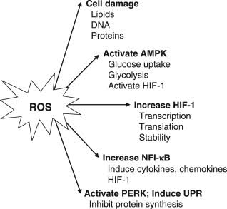
Reactive oxygen species are generated through both physiological and pathological mechanisms. Their production depends on cellular metabolism, oxygen availability, enzymatic activity, and environmental conditions.
Mitochondrial ROS Production
Mitochondria play a central role in energy metabolism through oxidative phosphorylation. During electron transport, a small proportion of electrons may escape the respiratory chain and interact with oxygen, generating superoxide anions.
Superoxide can subsequently be converted into hydrogen peroxide through the activity of superoxide dismutase enzymes. Hydrogen peroxide is relatively more stable and can participate in redox signaling or be converted into other reactive species.
Changes in mitochondrial function may alter ROS production and affect membrane potential, ATP synthesis, calcium regulation, and cellular survival.
Enzymatic Sources of Reactive Species
Several enzymes contribute to the generation of reactive oxygen and nitrogen species.
NADPH oxidases are particularly important because they produce ROS as part of regulated signaling and immune defense mechanisms. Xanthine oxidase can also contribute to superoxide and hydrogen peroxide production during purine metabolism.
Nitric oxide synthases generate nitric oxide from L-arginine. Under certain conditions, alterations in enzyme coupling may contribute to additional superoxide production, influencing the formation of reactive nitrogen species.
Environmental and Metabolic Influences
Cellular ROS production can also be influenced by external and metabolic factors, including:
Exposure to certain chemical compounds.
Inflammatory mediators.
Changes in oxygen availability.
Metabolic disturbances.
Ultraviolet and ionizing radiation.
Mitochondrial metabolic dysfunction.
Changes in intracellular calcium concentrations.
Investigating these sources helps researchers understand how different stress conditions influence cellular redox regulation.

3. Antioxidant Defense Mechanisms
Cells possess several complementary antioxidant systems that control reactive species and protect biological structures against excessive oxidation.
These defense mechanisms include enzymatic antioxidants, non-enzymatic molecules, and intracellular regulatory pathways.
Superoxide Dismutase
Superoxide dismutase (SOD) catalyzes the conversion of superoxide anions into hydrogen peroxide and molecular oxygen.
Three major forms are commonly described:
SOD1: Primarily located in the cytoplasm and also present in the mitochondrial intermembrane space.
SOD2: Located in the mitochondrial matrix.
SOD3: An extracellular antioxidant enzyme.
By controlling superoxide concentrations, SOD enzymes contribute to the regulation of oxidative reactions and mitochondrial function.
Catalase
Catalase is an antioxidant enzyme that converts hydrogen peroxide into water and molecular oxygen.
It is particularly abundant in peroxisomes, where it contributes to hydrogen peroxide regulation and helps prevent excessive accumulation of this reactive molecule.
Glutathione Peroxidase
Glutathione peroxidases reduce hydrogen peroxide and certain lipid hydroperoxides through reactions involving reduced glutathione (GSH).
During this process, glutathione is oxidized into glutathione disulfide (GSSG). The enzyme glutathione reductase subsequently regenerates reduced glutathione using NADPH.
The GSH/GSSG ratio is frequently investigated as an indicator of intracellular redox status, although its interpretation depends on the cellular compartment and experimental conditions.
Non-Enzymatic Antioxidants
In addition to enzymatic defenses, several small molecules contribute to antioxidant protection.
These include glutathione, ascorbic acid, vitamin E, and other endogenous or dietary compounds. Their mechanisms differ according to their chemical properties, cellular distribution, and interactions with reactive species.
The coordinated activity of these systems contributes to the maintenance of cellular redox homeostasis.
4. Molecular Pathways Involved in Cellular Stress Responses
Cells respond to oxidative challenges through interconnected signaling pathways that regulate antioxidant defenses, metabolism, inflammation, and survival.
The Nrf2 Signaling Pathway
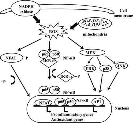
Nuclear factor erythroid 2-related factor 2 (Nrf2) is an important transcriptional regulator involved in cellular antioxidant responses.
Under basal conditions, Nrf2 is regulated by Kelch-like ECH-associated protein 1 (Keap1), which facilitates its degradation.
When oxidative or electrophilic conditions modify Keap1-dependent regulation, Nrf2 can accumulate and translocate into the nucleus. It then interacts with antioxidant response elements (AREs), promoting the expression of genes involved in cellular defense.
Important Nrf2-regulated targets include:
Heme oxygenase-1 (HO-1).
NAD(P)H quinone dehydrogenase 1 (NQO1).
Glutamate-cysteine ligase.
Glutathione-related enzymes.
Other proteins involved in detoxification and antioxidant regulation.
The Nrf2 pathway is therefore a major research target in investigations of oxidative adaptation and cellular protection.
The NF-κB Signaling Pathway
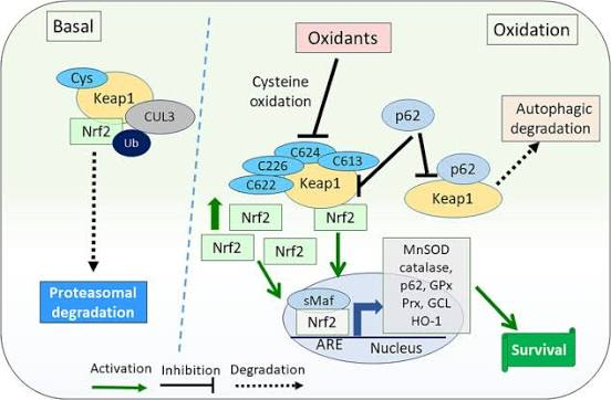
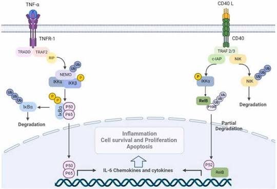
Nuclear factor kappa B (NF-κB) is a transcription factor involved in inflammatory regulation, immune responses, and cellular stress signaling.
Reactive species can influence NF-κB activity through several mechanisms, although the relationship is complex and dependent on cell type, stimulus, and intracellular context.
Following activation, NF-κB can regulate the expression of inflammatory mediators, cytokines, and proteins involved in immune responses.
Interactions between Nrf2 and NF-κB signaling are particularly relevant because antioxidant regulation and inflammation frequently influence one another.
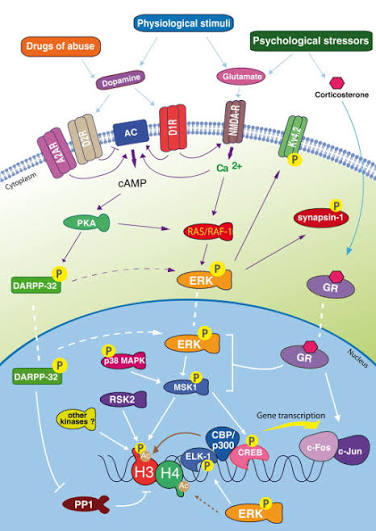
The MAPK Signaling Pathways
Mitogen-activated protein kinases (MAPKs) are involved in the transmission of extracellular and intracellular signals.
The principal MAPK pathways include:
ERK1/2: Frequently associated with cell proliferation, differentiation, and survival.
JNK: Involved in stress responses and apoptosis regulation.
p38 MAPK: Participates in inflammatory signaling, cellular adaptation, and stress-associated responses.
Oxidative changes can influence MAPK activity, contributing to modifications in gene expression and cellular behavior.
5. Oxidative Stress and Cellular Damage
When reactive species exceed the capacity of antioxidant defense mechanisms, oxidative stress can develop. Excessive oxidation may affect different classes of biological molecules and interfere with essential cellular functions.
Lipid Peroxidation
Cellular membranes contain polyunsaturated fatty acids that are particularly susceptible to oxidation.
Lipid peroxidation involves a chain reaction in which reactive species initiate the oxidation of membrane lipids. This process can generate secondary products, including malondialdehyde (MDA) and 4-hydroxynonenal (4-HNE).
These products are frequently investigated as indicators of oxidative damage and may influence membrane organization, protein function, and intracellular signaling.
Protein Oxidation
Reactive species can modify amino acid residues, alter protein structures, and affect enzymatic activity.
Oxidative modifications may result in protein carbonylation, changes in disulfide bonds, aggregation, or altered interactions with other cellular components.
Such changes can interfere with metabolic pathways and cellular regulatory mechanisms.
DNA and RNA Oxidation
Nucleic acids can also be affected by oxidative reactions.
One commonly investigated marker of oxidative DNA damage is 8-hydroxy-2'-deoxyguanosine (8-OHdG). Oxidative modifications of RNA may also influence RNA stability, translation, and gene expression.
Cells possess DNA repair and RNA quality-control mechanisms that help manage these modifications.
6. Mitochondrial Function and Oxidative Regulation
Mitochondria are essential regulators of cellular energy production, calcium homeostasis, and metabolic signaling. Their relationship with oxidative balance is particularly important because they are both sources and targets of reactive oxygen species.
Mitochondrial dysfunction may lead to changes in electron transport, membrane potential, and ATP production. These alterations can influence ROS generation and activate additional stress responses.
Mitochondrial quality-control mechanisms help maintain functional organelles through processes such as mitochondrial biogenesis, fusion, fission, and mitophagy.
Mitophagy is a selective form of autophagy responsible for the removal of damaged mitochondria. The PINK1/Parkin pathway is one of the best-characterized mechanisms involved in this process.
Research into mitochondrial redox regulation provides valuable information about cellular adaptation, metabolic dysfunction, and neurobiological mechanisms.
7. Oxidative Stress in Neurobiology
The nervous system has several characteristics that make oxidative regulation an important subject of investigation. Neurons have high metabolic requirements, depend heavily on mitochondrial energy production, and contain membrane structures rich in polyunsaturated fatty acids.
Oxidative imbalance has therefore been investigated in several neurological conditions, including Parkinson's disease, Alzheimer's disease, and other neurodegenerative disorders.
In Parkinson's disease research, dopaminergic neuronal models are frequently used to investigate oxidative mechanisms, mitochondrial dysfunction, and cellular responses to neurotoxic compounds.
Experimental models involving 6-hydroxydopamine (6-OHDA) provide opportunities to study oxidative damage, mitochondrial alterations, and neuronal cell viability.

Researchers may examine several complementary parameters:
Intracellular ROS production.
Mitochondrial membrane potential.
Antioxidant enzyme activities.
Glutathione concentrations.
Lipid peroxidation.
Inflammatory signaling.
Expression of genes involved in cellular stress responses.
Cell viability and membrane integrity.
These investigations help characterize the relationships between oxidative mechanisms and neuronal function without assuming that a single molecular pathway explains the entire disease process.
8. Experimental Methods for Evaluating Oxidative Balance
The investigation of oxidative stress requires complementary analytical approaches because no single biomarker provides a complete representation of cellular redox status.
Measurement of Intracellular ROS
Fluorescent probes such as 2',7'-dichlorodihydrofluorescein diacetate (DCFH-DA) are commonly used to investigate changes in intracellular oxidant-dependent fluorescence.
After entering cells, the probe undergoes intracellular deacetylation. Its subsequent oxidation produces a fluorescent signal that can be measured using fluorescence microscopy or plate readers.
However, DCFH-DA is not specific to a single ROS type. Experimental controls and complementary measurements are therefore essential for appropriate interpretation.
Antioxidant Enzyme Activity
SOD, catalase, and glutathione peroxidase activities can be measured using different spectrophotometric or enzymatic assay methods.
These measurements provide information about the activity of antioxidant defense systems under different experimental conditions.
Evaluation of Lipid Peroxidation
Malondialdehyde is commonly investigated as a product of lipid peroxidation.
The thiobarbituric acid reactive substances (TBARS) assay is frequently used for this purpose, although it presents limitations in specificity. More selective analytical methods can provide additional information about lipid oxidation products.
Nitric Oxide and Reactive Nitrogen Species
Nitric oxide-related activity can be investigated using several approaches, including the Griess reaction for nitrite measurement and fluorescent probes designed to detect nitric oxide-related signals.
The Griess assay measures nitrite rather than nitric oxide directly. This distinction is important when interpreting experimental findings.
Gene Expression Analysis
Reverse transcription quantitative polymerase chain reaction (RT-qPCR) can be used to investigate the expression of genes associated with antioxidant defense, inflammation, and cellular stress.
Frequently studied targets include NFE2L2, HMOX1, SOD1, SOD2, CAT, GPX1, and inflammatory mediators such as IL6.
Appropriate reference genes, validated primers, biological replicates, and suitable experimental controls are essential for reliable interpretation.
9. Cellular Adaptation and the Concept of Redox Signaling
Not all increases in reactive species indicate cellular damage. Under certain conditions, moderate changes in ROS production can activate adaptive mechanisms that improve cellular resistance to subsequent challenges.
This phenomenon is associated with redox signaling and, in specific experimental contexts, hormetic responses.
Cells can respond to mild stress by activating antioxidant genes, modifying metabolic activity, and improving certain defense mechanisms.
However, the biological outcome depends on the intensity, duration, and nature of the stressor, as well as the cell type and its physiological condition.
Distinguishing adaptive redox signaling from damaging oxidative stress is therefore an important objective in experimental research.
10. Emerging Perspectives in Oxidative Stress Research
New analytical and molecular technologies are expanding the possibilities for investigating oxidative regulation.
Advanced imaging approaches allow researchers to examine changes in cellular redox activity with improved spatial resolution. Genomic and transcriptomic analyses provide information about stress-associated gene expression, while proteomic approaches help characterize oxidative protein modifications.
Metabolomics can also reveal changes in metabolic pathways associated with redox regulation, energy production, and antioxidant metabolism.
Emerging research directions include:
Compartment-specific redox measurements.
Mitochondrial oxidative signaling.
Interactions between oxidative stress and inflammation.
Redox regulation in neurodegenerative processes.
Molecular mechanisms of cellular adaptation.
Advanced fluorescent biosensors.
Integration of multi-omics datasets.
Computational modeling of cellular stress networks.
These approaches are contributing to a more detailed understanding of the interactions between oxidative balance, cellular metabolism, and biological responses.
Conclusion
Oxidative balance is a fundamental component of cellular homeostasis, involving the coordinated regulation of reactive species, antioxidant defenses, and molecular signaling pathways.
Mitochondria, antioxidant enzymes, redox-sensitive transcription factors, and cellular quality-control systems collectively contribute to the regulation of oxidative responses. When these mechanisms become disrupted, oxidative stress may influence membrane integrity, protein function, nucleic acid stability, and cellular survival.
The integration of biochemical assays, molecular biology techniques, advanced imaging, and computational analysis provides valuable opportunities to investigate these mechanisms in different biological models.
Continued research into oxidative balance and cellular stress responses will contribute to a deeper understanding of cellular adaptation, inflammatory regulation, mitochondrial function, and the molecular processes involved in human disease.